
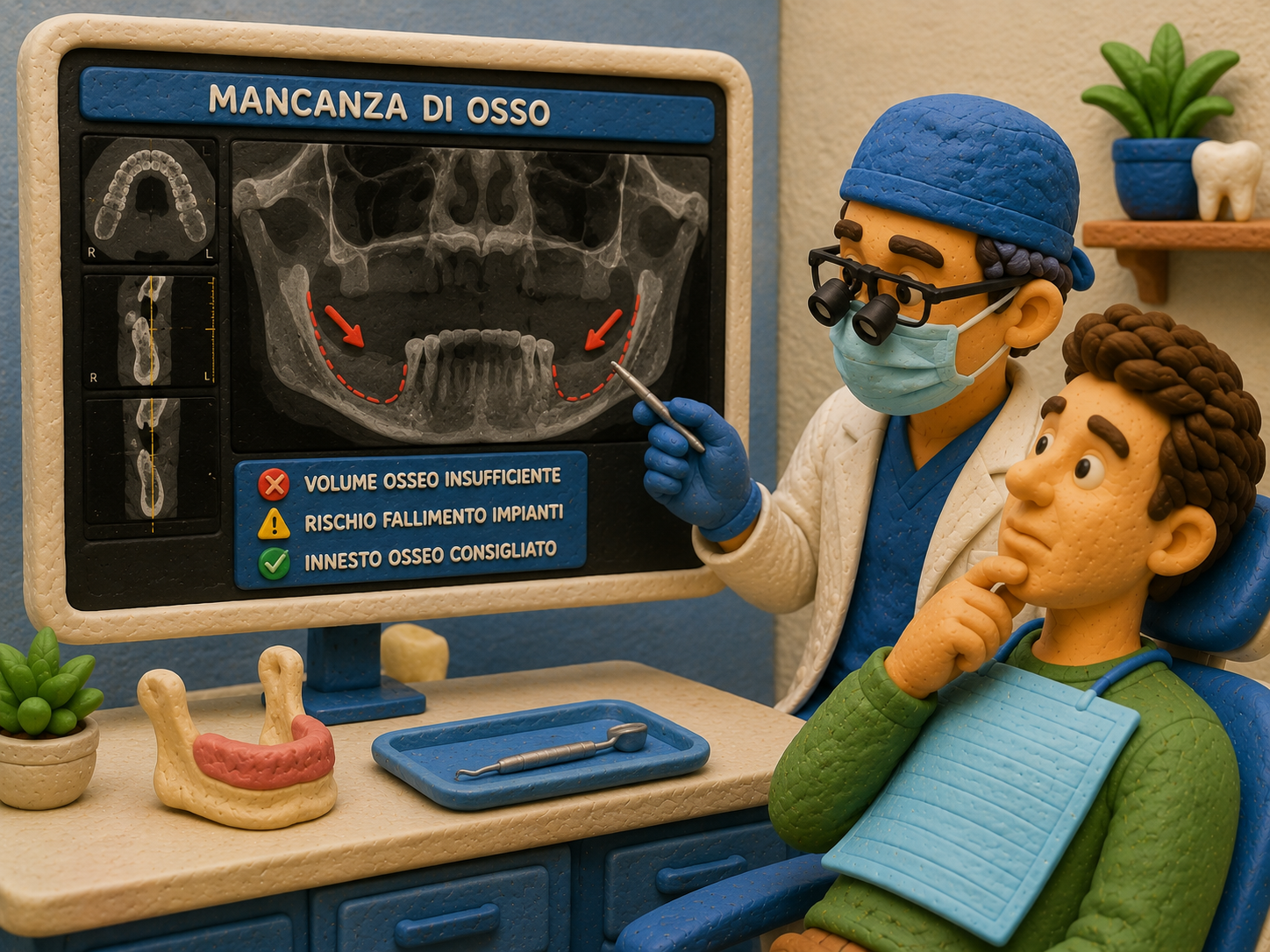
One of the most common obstacles on the way to a dental implant isn't the missing tooth itself, but what's left underneath it: the bone in the jaw or maxilla. When a tooth has been missing for a while, the bone that once supported it tends to resorb progressively — and if the remaining volume isn't sufficient, the implant has no solid base to integrate into.
In these cases, dentists often propose a bone graft: a procedure that rebuilds the missing volume before (or during) implant placement. Not all grafts are the same: different materials exist, with quite different characteristics, and the choice affects both the outcome and the healing timeline.
In this article
What bone atrophy is and why it happens
Jaw bone, like any bone tissue, stays healthy only if it is stimulated. When a natural tooth is present, chewing continuously transmits mechanical load to the root and from there to the surrounding bone, keeping it active. When the tooth is lost — through extraction, trauma, or periodontal disease — this stimulation stops, and the bone in that area begins to resorb: it loses volume in height and width, a process known as bone atrophy.
- Resorption is progressive: the more time passes after tooth loss, the more bone is lost, especially in the first 12-18 months.
- It isn't uniform: some areas of the jaw resorb faster than others, depending on the bone's original density.
- It has causes beyond simple extraction: advanced periodontal disease, prolonged use of poorly fitted removable dentures, trauma, infections.
- It's more pronounced in the upper jaw near the maxillary sinuses, where the bone is often already less dense to begin with.
Why an implant needs enough bone
A dental implant doesn't simply "rest" on the bone: it is inserted inside it and must integrate with it through a biological process called osseointegration, in which bone grows in direct contact with the titanium surface. For this to happen in a stable way, two things are needed together: sufficient height and width of healthy bone around the implant's entire length.
If the available bone is too thin or too short, the implant risks not achieving stable anchorage, becoming partially exposed over time, or, in the worst cases, failing to integrate at all. For this reason, before any implant — especially for full-arch rehabilitations such as All-on-4 or All-on-6 — the dentist assesses the actual bone volume with a cone-beam CT scan, not with a clinical exam alone.
A graft isn't always needed
Some techniques — such as the tilted implants used in All-on-4 — are specifically designed to make the best use of available bone and avoid a graft when resorption is mild or moderate. A graft is still necessary when the remaining bone, in height or width, falls below the minimum threshold for secure anchorage.
What a bone graft is and how it works
A bone graft (or guided bone regeneration) is a surgical procedure that places bone material in an area with insufficient volume, to stimulate the growth of new bone tissue from the patient around and within it. The grafted material works as a biological scaffold: over time, the patient's natural bone colonizes it and partly replaces it, increasing the volume and density of the area until it becomes suitable for an implant.
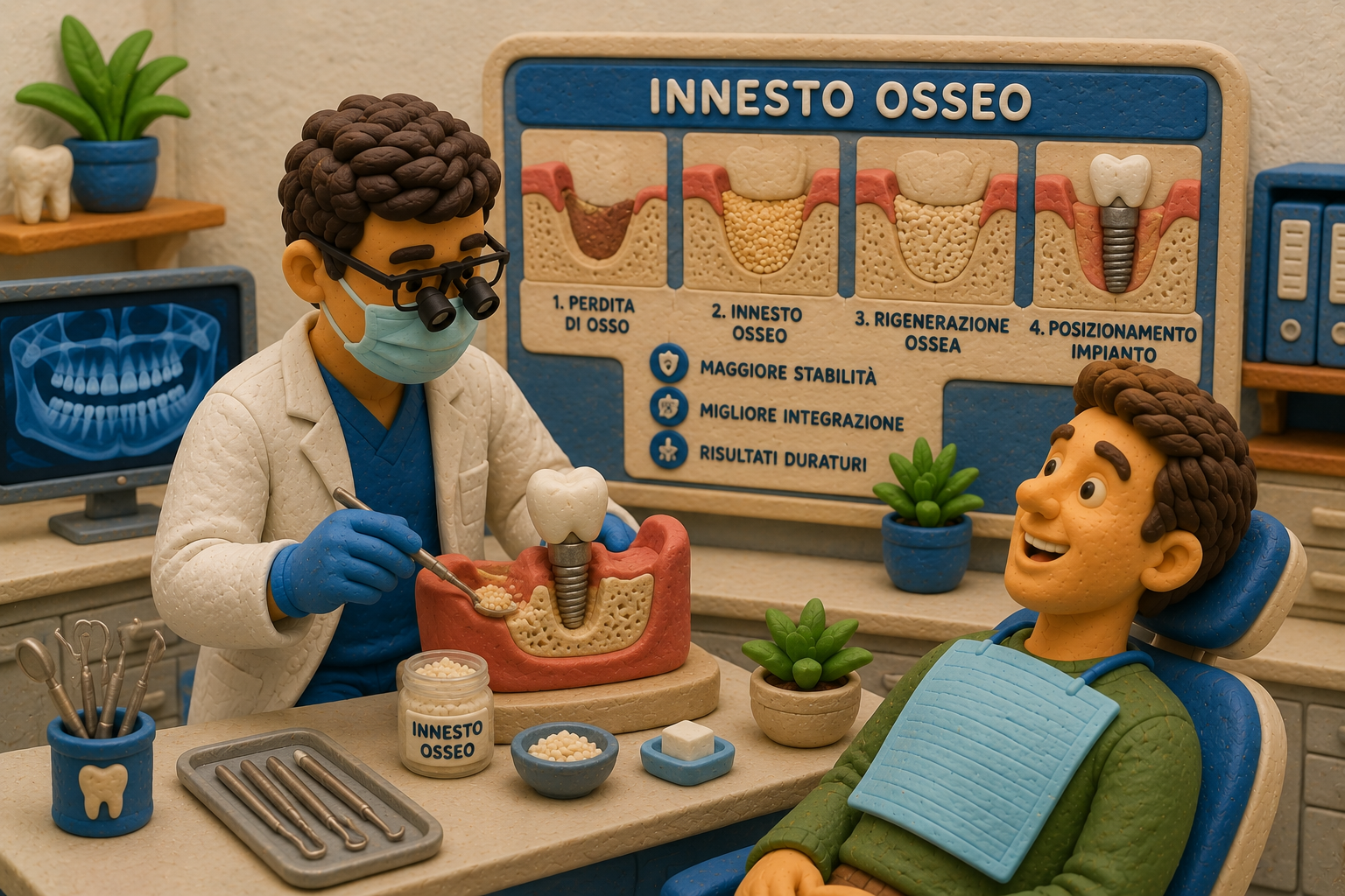
- Bone loss: the area to be treated has insufficient bone volume for a stable implant.
- Bone graft: the graft material is surgically placed in the area, often together with a protective membrane.
- Bone regeneration: over the following months, the patient's natural bone colonizes the grafted material, increasing its volume and density.
- Implant placement: once regeneration is complete (or, in some cases, during it), the implant is inserted into the rebuilt bone.
In some cases, when resorption is mild and the implant's initial stability is still assured, the graft and the implant can be performed in the same session. When the missing bone volume is more substantial, it is instead necessary to wait for the graft to heal before proceeding.
Types of graft material
Not all graft materials are equivalent: they differ in origin, regenerative capacity, and integration times. It's a point worth clarifying with your dentist before proceeding, because it affects both the outcome and, often, the price.
| Type | Origin | Things to consider |
|---|---|---|
| Autologous (autograft) | Taken from the patient's own body (chin, mandibular ramus, or, in extensive cases, hip or tibia) | Contains living cells and has the best regenerative capacity; requires a second surgical harvest site |
| Human donor (allograft) | Human bone from a tissue bank, processed and sterilized | Avoids a second surgical site while remaining human in origin; contains no living cells but provides an excellent biocompatible scaffold |
| Animal-derived (xenograft) | Usually processed and purified bovine bone | Widely used and well tolerated, but not of human origin; resorption is generally slower |
| Synthetic (alloplastic) | Lab-produced materials such as hydroxyapatite or tricalcium phosphate | No biological source tissue to consider, unlimited availability; regenerative capacity generally lower than human bone |
Why human bone remains the reference choice
Among these materials, bone of human origin is generally considered the best choice, because it is biologically closer to the tissue it needs to regenerate. Within that category, autologous bone — taken from the patient's own body — is the gold standard: it contains living cells and has the highest regenerative potential, at the cost of requiring a second harvest site. When taking bone from the patient isn't practical or necessary, human donor bone (allograft) is the best human-origin alternative, preferred by many clinicians over animal-derived or fully synthetic materials.
Timelines and healing
| Phase | Indicative duration |
|---|---|
| Graft surgery | 30 minutes to about 1-2 hours, under local anesthesia |
| Initial soft-tissue healing | 1-2 weeks |
| Bone regeneration (small, localized grafts) | 3-4 months |
| Bone regeneration (larger grafts, e.g. sinus lift) | 6-9 months |
| Implant placement | After regeneration, except in cases performed at the same time as the graft |
Timelines vary considerably depending on how much bone needs to be rebuilt, the material chosen, and individual healing capacity. A localized graft for a single tooth generally takes months, not weeks; an extensive sinus lift can take several more. Anyone considering treatment abroad should factor in that, when a graft is involved, the full pathway involves more than one trip — not just the one for the initial surgery.
Sinus lift: a special case
In the upper jaw, in the molar area, the bone borders the maxillary sinuses — two air-filled cavities within the facial bones. When the upper back teeth are lost, the bone in that area resorbs upward and the maxillary sinus tends to expand downward, further reducing the available bone space. This is one of the most frequent situations in which a graft is needed.
In these cases the procedure is called a sinus lift (or maxillary sinus augmentation): the membrane lining the sinus is gently lifted, and the space created underneath it is filled with graft material to rebuild the bone height needed for a stable implant. It's a well-established procedure, but more delicate and with a longer healing time than a graft localized to a single area.
- Lateral window sinus lift: indicated for more pronounced bone shortfalls, accessed through a small lateral opening.
- Crestal (osteotome) sinus lift: a less invasive technique, indicated when a smaller amount of bone is missing, often performed in the same session as the implant.
Risks, contraindications, and alternatives
- Infection or failed integration of the grafted material, which may require repeating the procedure.
- Post-operative swelling and discomfort, generally greater than with a simple implant, especially for extensive grafts.
- Longer overall timelines before reaching the final prosthesis.
- Factors that increase risk: smoking, uncontrolled diabetes, poor oral hygiene, systemic bone conditions.
A graft isn't always the only path: when resorption is mild or moderate, some implant techniques — such as All-on-4, which uses tilted posterior implants to anchor into denser areas of bone — often make it possible to avoid one. This is a case-by-case assessment based on the CT scan, not something decided in advance.
Questions to ask your dentist
- How much bone do I actually have available, according to the CT scan?
- What type of graft material are you proposing, and why is it right for my case?
- If donor or animal-derived bone is proposed, which tissue bank or supplier does it come from?
- Can the graft be avoided with an alternative technique, in my specific case?
- How much time should I expect between the graft and the final implant?
- Is the cost of the graft included in the quote, or priced separately after the CT scan?